PLANTAR FIBROMA
Plantar Fibroma: The Lump in the Arch That Isn't Plantar Fasciitis, Isn't a Cyst, and Usually Isn't an Emergency
A firm, slow-growing nodule inside the plantar fascia — the plantar fibroma is often found by accident, mistaken for a cyst, or blamed for pain that belongs to a different condition. Here is what it is at the tissue level, how to tell it apart from its neighbors, and the honest ladder of what to do about it.
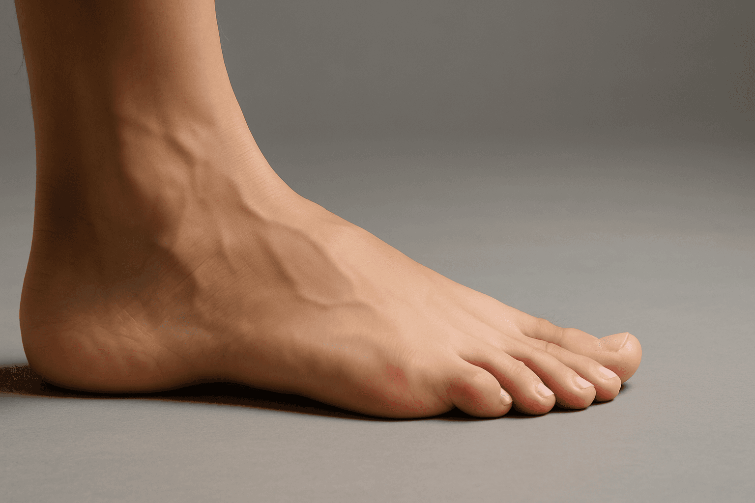
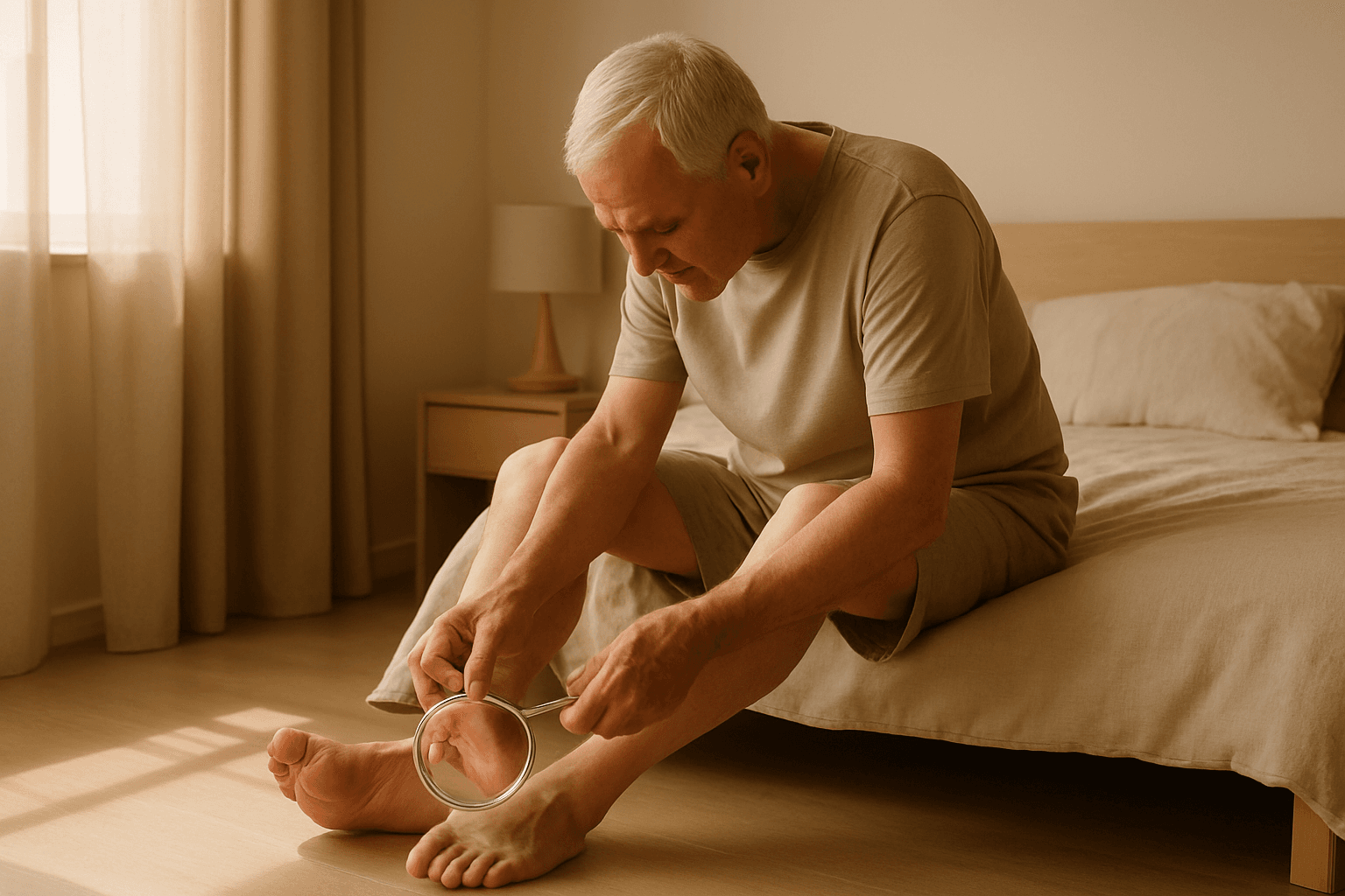
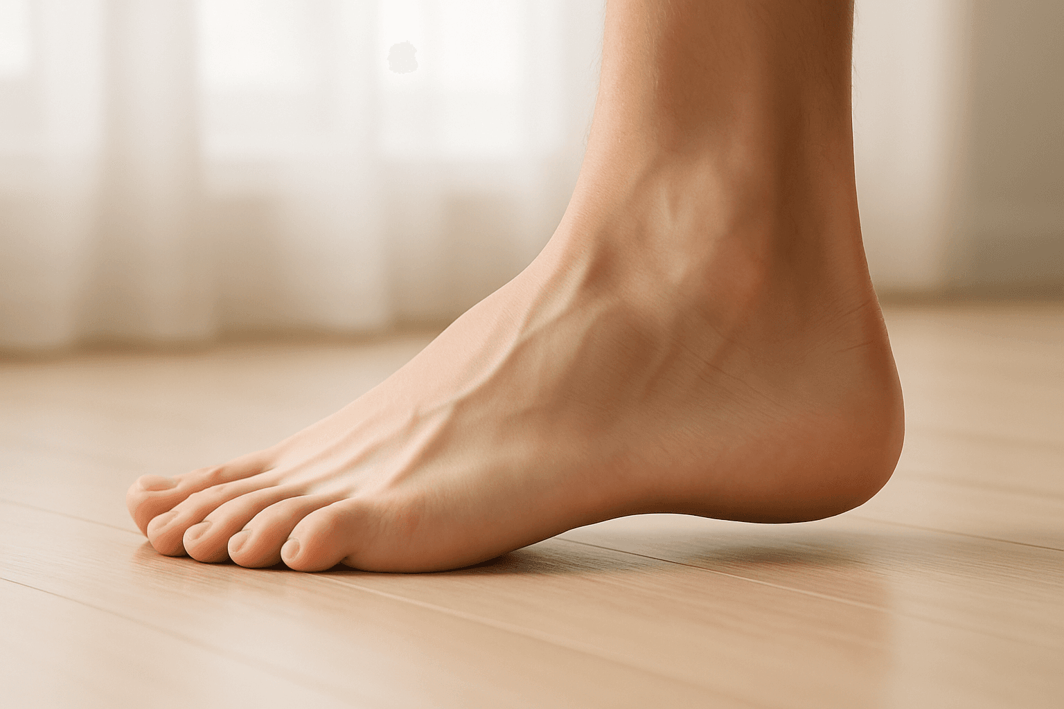
A plantar fibroma is a firm, slow-growing benign nodule that forms inside the plantar fascia — the thick band of tissue running from the heel bone to the base of the toes. Most readers find theirs by accident: a small, pea-sized bump in the arch of the foot that feels like a knot, that is noticed while washing in the shower or while putting on a sock, that may or may not hurt depending on the shoe. The nodule is a growth of fibroblasts and collagen within the fascia itself, part of a group of conditions called superficial fibromatoses that also includes Dupuytren's contracture of the hand and Peyronie's disease. It is not a cyst, not a cancerous tumor, and not plantar fasciitis — though it is often confused with each of those by patients and even by some non-foot-specialist clinicians.
This page is the walk-through we would want handed to us the morning after noticing a lump in the arch. It covers what the fibroma actually is at the tissue level, the specific features that tell it apart from the three conditions most readers worry it might be (a ganglion cyst, a soft tissue tumor, and the thickened fascia of plantar fasciitis), the common patterns of growth and symptoms, the honest conservative care that calms most symptomatic fibromas without surgery, the specific role of imaging and biopsy, and the surgical conversation that is reserved for a small subset of cases. For readers whose arch pain turns out to be plantar fasciitis after all, our bottom of heel pain walk-through and the full plantar fasciitis hub are the right next reads.
Nothing on this page is medical advice. A new lump in the foot — including one as benign as the typical plantar fibroma — deserves a hands-on exam by a podiatrist, orthopedist or primary care physician, both to confirm the diagnosis and to rule out the small list of conditions that can mimic a fibroma. Every recommendation on this hub is written for comfort, support and relief — nothing on this site is a treatment or a cure for a plantar fibroma.
What a plantar fibroma actually is
At the tissue level, a plantar fibroma is a nodular proliferation of fibroblasts (the cells that produce connective tissue) and the collagen fibers they lay down, growing within the plantar fascia itself rather than on top of or underneath it. Pathologists call the condition plantar fibromatosis or Ledderhose disease, after the German surgeon who described it in the nineteenth century. Under the microscope, a fibroma shows dense bundles of collagen with fibroblasts and myofibroblasts scattered through them — a tissue pattern similar to a scar or a healing tendon, but growing where there was no injury to repair.
The useful mental model for most readers is that a fibroma is a knot of the fascia's own material that formed where no knot should be. It is firm (the texture of a dense rubber eraser, not the softness of a fluid-filled cyst), it is fixed to the fascia (it does not slide independently under the skin the way a lipoma or a cyst often will), and it grows slowly over months to years. Fibromas are most commonly found along the medial band of the fascia, roughly in the middle of the arch, though they can appear anywhere along the fascia's length. The classic case is a single nodule between a quarter inch and an inch across; roughly a third of patients have multiple nodules, and roughly a quarter have fibromas in both feet.
Plantar fibromatosis belongs to a family of superficial fibromatoses that run in some families and are associated with a few specific conditions. The best-known relatives are Dupuytren's contracture (the same pattern of nodular fascial growth in the palm of the hand) and Peyronie's disease (in the penis); readers with any of those conditions in themselves or close family members are at higher risk of a plantar fibroma. The condition is also associated with diabetes, chronic alcohol use, epilepsy medications (specifically phenytoin), and some patterns of long-term microtrauma to the foot. Idiopathic cases — no family history, no risk factor, no injury — are common; a fibroma on its own in an otherwise healthy foot is a routine finding.
How to tell a fibroma apart from its lookalikes
The three conditions that most commonly get confused with a plantar fibroma are a ganglion cyst, a lipoma, and the thickened fascia of plantar fasciitis. The useful distinguishing features are different for each, and most of them can be read by a careful hands-on exam before any imaging. A ganglion cyst is a fluid-filled sac that typically arises from a joint or tendon sheath. It feels softer than a fibroma, often has a slight give when pressed, usually transilluminates when a light is shone through it in a dark room (because the fluid is clear), and sometimes shrinks and grows over weeks as the fluid volume changes. A fibroma is firm, does not transilluminate, and does not change size over short periods.
A lipoma is a benign tumor of fat cells, soft and compressible, that typically sits in the fat layer under the skin rather than inside the fascia. It usually slides independently under the skin when pressed from different angles, in a way a fibroma does not — the fibroma is anchored to the fascia and moves with it, which is often the clearest clinical finding on exam. A soft tissue sarcoma — the rare malignant lookalike that every clinician will mentally rule out — is usually larger, grows faster, and often has a different texture; the imaging workup for an unclear case usually settles the question before any biopsy.
Plantar fasciitis is the diagnosis most readers worry a fibroma might really be, often because the arch pain and the fascia location overlap. The useful distinction is that plantar fasciitis pain is at the fascia's heel attachment (the medial heel) and is worst with the first steps in the morning, while a fibroma is usually in the middle of the arch and is more consistently painful when a shoe or a surface presses directly on it rather than following a morning-first pattern. A reader can have both at once — a thickened, inflamed fascia at the heel plus one or more fibromas in the arch — and the diagnoses are compatible rather than competing. Our bottom of heel pain walk-through covers the plantar fasciitis pattern in detail.
How a fibroma usually behaves
The common story is a reader in their forties or fifties noticing a small firm bump in the arch of one foot, usually without a specific inciting event. The nodule is a quarter to a half inch across on first notice, is not painful on its own, but hurts if a shoe seam, a floor, or a running insole presses directly on it. Over the following months to years, the fibroma typically either stays the same size, grows slowly, or occasionally shrinks on its own; rapid growth is uncommon and is the one feature that moves a case up the diagnostic urgency ladder. Many fibromas remain asymptomatic indefinitely and are found incidentally during a foot exam for an unrelated problem.
The symptom pattern is almost entirely a pressure story. A fibroma that sits in a part of the arch where no shoe presses hurts almost not at all; the same fibroma in a tight dress shoe or under a firm-arched insole can be sharply tender with each step. Barefoot walking on a hard floor often brings the pain out, especially on tile or concrete, because the floor presses directly on the nodule. Soft surfaces (grass, carpet, a cushioned shoe) are usually comfortable regardless of the fibroma's size. The useful implication is that most of the daily discomfort from a fibroma is a shoe and insole problem rather than a tissue problem, and the first useful intervention is almost always a cushioning accommodation around the nodule rather than any direct treatment of the fibroma itself.
Over longer timelines (months to years), a growing fibroma can occasionally reach a size where it pushes on surrounding structures, affects gait, or produces a visible deformity of the arch. These cases are the minority; most fibromas plateau at a size that remains a shoe-fit nuisance rather than a mechanical problem. Rapid growth, severe pain, numbness or tingling radiating into the toes, or any change in the color or temperature of the surrounding skin is out of the typical fibroma pattern and deserves a prompt in-person exam.
What imaging and biopsy actually add
The initial diagnosis of a plantar fibroma is often clinical — a podiatrist or foot-and-ankle orthopedist with a hands-on exam can usually read a typical fibroma without any imaging. The useful role of imaging is to confirm the diagnosis in less typical cases, map the exact extent of the nodule if surgery is being discussed, or rule out the small list of lookalikes. Ultrasound is usually the first imaging test ordered: it is cheap, fast, and shows a fibroma's characteristic hypoechoic (dark) nodule within the fascia, with the surrounding fascia visible for comparison. Magnetic resonance imaging is a second step for cases where ultrasound is unclear, where surgery is being planned, or where a soft tissue sarcoma needs to be ruled out — MRI shows the fibroma, its connection to the fascia, and the surrounding tissue in detail.
A biopsy is rarely needed and is usually only ordered when the clinical and imaging features are atypical enough to raise concern about a different diagnosis. The useful indicators that push toward biopsy are a nodule that grew unusually fast, that is unusually large (over an inch and a half across), that has imaging features that do not fit a typical fibroma, or a patient with a personal history of a different soft tissue tumor. For a typical fibroma with a classic exam and ultrasound, biopsy is not usually indicated — the imaging has already answered the question with higher certainty than most patients expect.
Pathology, when it is done, confirms the diagnosis definitively by showing the collagen-and-fibroblast tissue pattern. The main reason some surgeons send a resected fibroma to pathology after a surgical removal (rather than before) is to confirm the diagnosis after the fact rather than to drive treatment choice; the surgery itself is usually planned on the clinical and imaging picture.
The conservative care ladder for a symptomatic fibroma
Most symptomatic plantar fibromas respond well to a conservative care ladder that focuses on accommodation rather than removal. The first and most useful move is a shoe-and-insole change that stops pressing directly on the nodule. The specific accommodation is often a custom insole with a cut-out or depression where the fibroma sits, so the surrounding arch is supported while the nodule itself floats in a soft pocket. Many podiatrists make these as a quick in-office modification to an off-the-shelf insole; a trial-and-error approach using a plain cushioned insole and a small adhesive gel pad placed around (not over) the nodule often works as a first step. Softer, better-cushioned shoes with more room in the midfoot are a parallel useful change, and barefoot walking on hard floors is worth minimizing. Our how to use heel cups walk-through covers the related insole accommodation decisions.
Topical treatments and physical therapy sit at the second rung. Daily stretching of the plantar fascia and the Achilles complex — the same routine covered in our plantar fasciitis stretches walk-through — is often recommended to maintain fascia elasticity and reduce the tension that may contribute to nodule growth; the evidence for stretching slowing a fibroma itself is thin, but the broader fascia health is net positive. Topical verapamil cream (a calcium channel blocker compounded for topical use) has a small body of evidence for softening and slowly shrinking plantar fibromas over several months of nightly application; it requires a prescription and a compounding pharmacy. Topical NSAID gels help with pain around a tender nodule but do not change the fibroma itself.
Corticosteroid injections directly into the fibroma are a third rung and have mixed evidence. Some case series show modest shrinkage and symptom improvement; others show little effect and raise concern about surrounding fat pad atrophy if injections are repeated. The decision to inject is a conversation with a foot specialist based on the specific nodule, its symptoms, and the patient's broader risk tolerance. The fourth rung — reserved for a smaller group — is extracorporeal shockwave therapy, which has a growing but still limited evidence base for Ledderhose disease and is sometimes offered at specialist clinics. Radiation therapy, used routinely for Dupuytren's contracture in the hand in some countries, is less commonly offered for plantar fibromas in the United States but is a real option at some centers.
When surgery enters the conversation
Surgical removal of a plantar fibroma is reserved for cases that have failed six to twelve months of honest conservative care, that are causing severe pain or genuine difficulty walking, or that have grown rapidly enough to raise concern about the diagnosis itself. The operation is not a small one, and the recurrence rate — the fibroma coming back at the same or a nearby site — is high enough that most foot specialists are reluctant to recommend surgery for anything short of a case that has clearly failed conservative management.
The two main surgical approaches are a local excision (removing the nodule itself with a small margin of surrounding fascia) and a wider fasciectomy (removing a longer segment of the fascia along with the nodule). Local excision has shorter recovery and lower surgical risk, but a reported recurrence rate of thirty to fifty percent in published series. Wider fasciectomy has a lower recurrence rate (reports range from ten to twenty-five percent) but longer recovery, more surgical risk, and the biomechanical consequence of removing a chunk of a structural tissue. For most patients, the local excision is the first operation; a recurrence after a local excision sometimes prompts a wider procedure, though many patients choose to go back to conservative accommodation instead.
Recovery from either operation involves roughly two to four weeks in a surgical shoe or walking boot, then a gradual return to normal footwear and activity over the following six to twelve weeks. Full fascia healing takes several months, and the surgical scar itself can be sensitive and tender for longer than that. Patients considering surgery should have an honest conversation with their foot specialist about the recurrence risk, the recovery timeline, and the biomechanical change in the arch after the operation. For most patients with a stable, modestly symptomatic fibroma that responds to shoe accommodation, surgery is a step too far; for the small group with severe pain or a nodule that is clearly affecting gait, it is a reasonable option with eyes open to the trade-offs.
Nothing on this page replaces a hands-on exam. A new lump in the foot, a growing lump that has not been evaluated, or a lump associated with new pain, numbness, swelling, warmth or color change deserves an in-person visit with a podiatrist, orthopedist or primary care physician. The useful rule is that most foot lumps are benign, that most benign foot lumps are stable or slow-growing fibromas, cysts or lipomas, and that the small list of serious lookalikes is almost always sorted out by a careful exam and a single ultrasound.
The full heel pain map, with the plantar fasciitis differential
A lump in the arch is one of several patterns that get labeled as plantar fasciitis before an exam. The heel pain hub walks through the full differential, where the fibroma fits inside it, and the daily routine for the plantar fasciitis cases that often sit alongside it.
The full heel pain map, with the plantar fasciitis differential →Frequently asked questions
Is a plantar fibroma dangerous or cancerous?
A plantar fibroma is a benign growth of fascia tissue and does not become cancerous. It is one of the superficial fibromatoses, a family of conditions that also includes Dupuytren's contracture of the hand. The small list of concerning lookalikes — a soft tissue sarcoma most notably — is almost always sorted out by a careful exam and a single ultrasound, which is why any new lump in the foot deserves a hands-on evaluation even when the clinical picture is reassuring. Signs that would move a case up the urgency ladder include rapid growth, a size above an inch and a half across, severe or constant pain, or associated numbness, tingling, swelling or color change in the surrounding skin.
What causes a plantar fibroma?
The precise cause is unknown. The condition has a clear hereditary component — close relatives of patients with plantar fibromatosis, Dupuytren's contracture or Peyronie's disease are at higher risk — and shares some genetic signatures with those conditions. Associated factors include diabetes, long-term alcohol use, phenytoin and some other epilepsy medications, and some patterns of long-term microtrauma to the plantar fascia. Many cases are idiopathic (no family history, no associated condition, no clear trigger), and a fibroma on its own in an otherwise healthy foot is a routine clinical finding.
Does a plantar fibroma go away on its own?
Most plantar fibromas do not resolve spontaneously once they have formed. The common natural history is a slow growth to a stable size, followed by months to years of relatively unchanged size. Spontaneous shrinkage is reported occasionally in case series but is not something to count on. The useful implication is that most symptomatic fibromas are managed with accommodation and ongoing care rather than waited out, and that a fibroma that is asymptomatic is often best left alone and monitored rather than treated for its own sake.
What is the best treatment for a plantar fibroma?
For most symptomatic fibromas, the useful first move is a shoe-and-insole accommodation that stops pressing directly on the nodule — a custom or modified insole with a cut-out or depression where the fibroma sits, paired with a softer, better-cushioned shoe. Daily plantar fascia and calf stretching, topical verapamil cream (prescription, compounded), and occasionally a corticosteroid injection are the next rungs. Extracorporeal shockwave therapy is a specialist option at some clinics. Surgical excision is reserved for cases that have failed six to twelve months of honest conservative care and that are causing severe pain or genuine difficulty walking; the recurrence rate after surgery is meaningfully high, which is why most foot specialists are reluctant to recommend surgery for mild or moderate cases.
Can I still walk and exercise with a plantar fibroma?
Most patients with plantar fibromas can walk and exercise normally, with adjustments. The useful rule is to protect the specific area around the nodule from direct pressure — a cushioned shoe, an insole with a cut-out or soft pocket around the fibroma, and avoiding barefoot walking on hard floors is usually enough to let normal activity continue. Running, hiking and standing jobs are generally fine with the right shoe accommodation; the limiting factor is almost always the shoe fit around the nodule rather than the exercise itself. A fibroma that is causing pain with activity even in a well-fitting shoe is a reason to see a foot specialist for a more targeted accommodation or a conversation about the next rung of care.
How is a plantar fibroma different from plantar fasciitis?
The two conditions involve the same tissue but in different ways and at different locations. Plantar fasciitis is an irritation and thickening of the plantar fascia at its heel attachment, producing the classic morning stab under the medial heel that eases with the first steps. A plantar fibroma is a discrete firm nodule in the fascia itself, usually in the middle of the arch, that is tender when pressed on but does not follow the morning-first pattern. A reader can have both at the same time, and the diagnoses are compatible rather than competing — a thickened heel attachment of the fascia plus one or more fibromas in the arch is a routine combination. The treatment ladders are different: plantar fasciitis responds to stretching, supportive insoles and load management; a plantar fibroma responds to accommodation around the nodule and, in a smaller group, the specialist ladder covered above.
HeelCups is reader-supported. When you buy through links on our site we may earn an affiliate commission, at no extra cost to you. This article is general information about foot comfort and is not medical advice — see our medical disclaimer.